Agriculture
 The Chrysophyceae, classified within the kingdom Chromista, are mostly unicellular or colonial organisms found in fresh and salt water throughout the world.
The Chrysophyceae (in some systems corresponding to the phylum Chrysophyta) are related to heterokont algae and include more than eight hundred described species that are classified in approximately one hundred genera.
They aremost closely related to the Synurophyceae and other pigmented heterokont algae, including the Bacillariophyceae (diatoms), Eustigmatophyceae, Phaeophyceae (brown algae), and Xanthophyceae (yellow-green algae), among others.
The classification of chrysophycean species remains in a state of flux. In one system of classification primary importance is placed upon the number of flagella (zero, one, or two) that are present in the motile cell stage.
A second classification organizes species based upon the predominant vegetative state of the organism. For example, in this classification amoeboid, coccoid, palmelloid, and flagellate species are assigned to separate orders.
Ecology and Diversity
Chrysophytes are predominantly found in fresh-water environments, although some are marine, and a few are reported from soil or snow.
 Members of the group are widely distributed but are most common in cold-temperate lakes, ponds, bogs, and ditches. Some species are common members of the phytoplankton,whereas others are epibionts or are neustonic (attached to the surface film of quiet water). Other species are only rarely observed.
Most chrysophytes are free-swimming unicellular or colonial flagellates. Others are coccoid (that is, immobile, walled unicells), amoeboid, or palmelloid (with cells enveloped in a gelatinous matrix). A few species are parenchymatous.
Cell Walls
Most chrysophytes lack a cell wall, but others produce species-specific outer coverings of scales or loricae. For example, complex siliceous scales or spines that are produced in silica deposition vesicles cover the cells of Paraphysomonas. The scales of Chrysolepidomonas are organic and of two types: those that are dendritic (tree-shaped) and those that are canistrate (cylindrical).
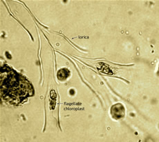
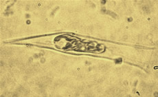
The cells of other species may be enclosed within an organic vaselike or flasklike lorica composed of cellulose and proteins or chitin (for example, Dinobryon, Pseudokephyrion, Poteriochromonas, Lagynion, and Stenocalyx).
 In such species the lorica is typically composed of fine, interwoven fibrils. In Dinobryon these fibrils are helically arranged and secreted as the cell rotates about its longitudinal axis.
In contrast, the loricae of Epipyxis species are composed of imbricate, overlapping scales. The posterior pole of the cell is typically positioned at the base of the lorica and may be attached by a fine cytoplasmic extension; the flagella protrude externally through the lorica opening.
Flagella
Chrysophytes are heterokont, biflagellate organisms that swim with at least one flagellum forwardly directed. The two flagella ofmotile cells are anteriorly inserted in an apical or subapical position and are unequal in length.
The flagella differ morphologically and are heterodynamic. In most species, the basal bodies from which the flagella arise are either oriented at an acute angle to one another or are perpendicular to one another.
In Hydrurus, Chromphyton, and Lagynion, the basal bodies form an obtuse (oblique) angle with respect to one another. The long (immature) flagellum is anteriorly directed and is ornamented with two rows ofmastigonemes and finer lateral filaments. Eachmastigoneme is composed of a base, a tubular shaft, and one to three terminal filaments; these are known as tripartite tubular hairs.
Mastigonemes are produced in the perinuclear space between the two outer membranes of the chloroplast and the two surrounding membranes of the chloroplast endoplasmic reticulum. The long flagellum beats in an undulatory, sine-wave-like motions that are initiated at the base of the flagellum.
The relatively stiff short (mature) flagellum is directed laterally or posteriorly, lacks mastigonemes, and rotates helically. A distinct swelling associated with the eyespot is typically present at the proximal base of the smooth flagellum.
In some taxa (such as Chromulina, Chrysococcus, and Sphaleromantis), the short flagellum is highly reduced and may be nonemergent; it is therefore undetectable by light microscopy. In a handful of species the short flagellum is entirely absent, although the mature basal body may persist within the cell.
 Naked motile cells bearing two visible flagella are often referred to as Ochromonas-like (or ochromonadalean), whereas those with one visible flagellum are typically assigned to the genus Chromulina.
The transitional region between the basal body and flagellum contains an electron-dense transitional plate, above which lies a coiled, apparently springlike transitional helix. The functions of the transitional plate and helix, which are also found in other flagellates, are uncertain.
In heterotrophic and mixotrophic species, the flagella play a role in prey capture. Particles actively captured by the flagella that are recognized as food are pushed into a feeding basket; those not recognized as food are released.
The feeding basket is formed and closed by movements of underlying microtubules (see below). Water currents produced by the undulation of the long flagellum may passively bring food particles in contact with the cells that, in some species, are collected by pseudopodia.
Cell Organization
Cells possess a single pear-shaped nucleus that is positioned at the anterior end of the cell. The narrow end of the nucleus typically lies close to the basal bodies. A prominent Golgi apparatus with distended cisternae lies against the nucleus. Contractile vacuoles (absent in some marine forms) are also found at the anterior end of the cell.
One or more mitochondria with tubular cristae are present in the cell. Because the mitochondria are usually long and coiled, the actual number of mitochondria present is difficult to discern. Fibrous bands, sometimes referred to as connecting fibers, connect the basal bodies to one another.
A cross-striated band of fibers known as the rhizoplast extends from the basal apparatus and forms a connection to the nucleus. Typically four microtubular roots (R1, R2, R3, and R4) originate near the basal bodies, take characteristic paths through the cell, and proliferate beneath the plasmalemma.
For example, in most species roots R3 and R4 often form a loop beneath the short flagellum. Other microtubules are nucleated from the four major roots that provide the cytoskeletal elements needed to maintain cell shape.
Muciferous bodies or discobolocysts are present in some species. Muciferous bodies are capable of extruding long threads, whereas discobolocysts forcefully eject discoid projectiles. These functions of these organelles have been little studied but may be involvedin prey capture or predator avoidance.
Nutrition
The Chrysophyceae employ a variety of means to obtain energy. Most chrysophytes are photosynthetic but require an exogenous source of vitamins (such as vitamin B12, biotin, and thiamin) for growth.
It is probable that all chrysophytes are opportunistically or facultatively osmotrophic; that is, they are capable of directly absorbing small inorganic or organic molecules (such as sugars and amino acids) from the surrounding medium. Several species, particularly those with leucoplasts, are obligate heterotrophs that are bactivorous or consume small organic particles.
Mixotrophic species are also well represented among the chrysophytes. This category includes photosynthetic species that, routinely or under unfavorable conditions, supplement their nutrition via phagotrophy.
Chloroplasts, Photosynthetic Pigments, and Storage Products
The chloroplasts of chrysophytes are typically golden-brown or yellow-green in color, and there are usually one to two chloroplasts per cell.
Chloroplasts are peripherally located, and pyrenoids may be present or absent. Four unit membranes surround each chloroplast; the outer two are derived from the endoplasmic reticulum and are typically continuous with the nuclear envelope.
Chloroplast lamellae are typically composed of three adpressed thylakoid membranes, and a girdle lamella, which completely encircles the chloroplast, is usually present. The chloroplast deoxyribonucleic acid (DNA) is ring-shapedandlies just beneath the girdle lamella.
The light-harvesting complex of chrysophytes contains chlorophylls a and c, beta-carotene, and the xanthophylls fucoxanthin, neoxanthin, violaxanthin, and zeaxanthin. Among these, fucoxanthin is dominant and is therefore responsible for the golden-brown color observed in most chrysophytes.
The major product of photosynthesis is a water-soluble ?-1,3-linked glucan (known as chrysolaminarin or leucosin) that is stored in cytoplasmic vacuoles in the posterior region of the cell. Lipids may also be produced and are also stored in the cytoplasm.
Eyespots (or stigmata) are present in many, but not all, species. The eyespot takes the form of a single layer of orange or reddish colored, lipidlike droplets that are located just beneath the chloroplast membrane. These droplets lie near a swelling located at the base of the smooth (short) flagellum; together the eyespot and flagellar swelling form a photoreceptor apparatus.
Several chrysophyte genera are known that contain a vestigial chloroplast (leucoplast) that lacks pigments (including Anthophysa, Monas, Oikomonas, Paraphysomonas, and Spumella).
Reproduction
Asexual reproduction in amoeboid and flagellate species occurs by longitudinal division of the cell; fragmentation is common among colonial, palmelloid, and parenchymatous species. In coccoid species reproduction may proceed via cell division or the formation of autospores that rupture and exit the parent cell wall.
Some taxa, such as the parenchymatous genera Phaeodermatium and Hydrurus or members of the palmelloid family Chrysocapsaceae, reproduce by means of flagellated swarmers (zoospores).
Under certain environmental conditions, silicified resting cysts, or statospores, are produced by many species. Statospores are formed endogenously, are roughly spherical or ellipsoidal, and have walls that may be smooth or ornamented.
The stomatocyst opening (porus) may be simple, possess a thickened collar, or take the form of a narrow neck. The cyst wall is formed by the deposition of silicate on an internal membrane, and the porus is preformed or produced by resorption of a portion of the cyst wall.
Depending on the species, cytoplasm located outside the cyst wall may or may not be absorbed through the porus, which at maturity is occluded by a pectic plug. During excystment the plug is lost, and one or more amoeboid or free-swimming flagellate cells emerge.
Sexual reproduction is known only in a handful of species. In those cases observed, vegetative cells behave as gametes and fuse apically. The resulting quadri flagellate cell (planozygote) will encyst forming sexually derived binucleate hypnozygotes or stomatocysts.
It is presumed that karyogamy (nuclear fusion) and meiosis occur within the cyst, but these processes have yet to be studied. Depending upon the species, sexual stomatocysts may give rise to one, two, or four vegetative cells.
- Cryptomonads
CryptomonadsThe phylum Cryptophyta describes tiny, motile, unicellular organisms with two slightly unequal flagella bearing lateral hairs. Cryptomonads live mainly in marine and freshwater environments. Some cryptomonads are alga-like, with bluegreen,...
- Diatoms
DiatomsDiatoms are unicellular microorganisms of the phylum Bacillariophyta that are abundant in aquatic, semi aquatic, and moist habitats throughout the world, growing as solitary cells, chains of cells, or members of colonies. Diatoms, algal organisms...
- Green Algae
Green AlgaeThe green algae are a diverse group of eukaryotic organisms classified in the phylum Chlorophyta. They are considered eukaryotic because individual cells possess a prominent structural feature known as a nucleus, which houses the chemicals...
- Haptophytes
HaptophytesThe algal phylum Prymnesiophyta, or Haptophyta, is a monophyletic taxon that contains two hundred to three hundred extant species that are 4-40 microns in size. The phylum Haptophyta is divided into two subclasses, the Prymnesiophycidae and...
- Heterokonts
HeterokontsHeterokonts are a group of closely related phyla with flagella in pairs, one long and one short. They include oomycetes, chrysophytes, diatoms, and brown algae. The term ?heterokont? refers either to the flagellar arrangement of biflagellate...
Agriculture
Chrysophytes

The Chrysophyceae (in some systems corresponding to the phylum Chrysophyta) are related to heterokont algae and include more than eight hundred described species that are classified in approximately one hundred genera.
They aremost closely related to the Synurophyceae and other pigmented heterokont algae, including the Bacillariophyceae (diatoms), Eustigmatophyceae, Phaeophyceae (brown algae), and Xanthophyceae (yellow-green algae), among others.
The classification of chrysophycean species remains in a state of flux. In one system of classification primary importance is placed upon the number of flagella (zero, one, or two) that are present in the motile cell stage.
A second classification organizes species based upon the predominant vegetative state of the organism. For example, in this classification amoeboid, coccoid, palmelloid, and flagellate species are assigned to separate orders.
Ecology and Diversity
Chrysophytes are predominantly found in fresh-water environments, although some are marine, and a few are reported from soil or snow.

Most chrysophytes are free-swimming unicellular or colonial flagellates. Others are coccoid (that is, immobile, walled unicells), amoeboid, or palmelloid (with cells enveloped in a gelatinous matrix). A few species are parenchymatous.
Cell Walls
Most chrysophytes lack a cell wall, but others produce species-specific outer coverings of scales or loricae. For example, complex siliceous scales or spines that are produced in silica deposition vesicles cover the cells of Paraphysomonas. The scales of Chrysolepidomonas are organic and of two types: those that are dendritic (tree-shaped) and those that are canistrate (cylindrical).
The cells of other species may be enclosed within an organic vaselike or flasklike lorica composed of cellulose and proteins or chitin (for example, Dinobryon, Pseudokephyrion, Poteriochromonas, Lagynion, and Stenocalyx).

In contrast, the loricae of Epipyxis species are composed of imbricate, overlapping scales. The posterior pole of the cell is typically positioned at the base of the lorica and may be attached by a fine cytoplasmic extension; the flagella protrude externally through the lorica opening.
Flagella
Chrysophytes are heterokont, biflagellate organisms that swim with at least one flagellum forwardly directed. The two flagella ofmotile cells are anteriorly inserted in an apical or subapical position and are unequal in length.
The flagella differ morphologically and are heterodynamic. In most species, the basal bodies from which the flagella arise are either oriented at an acute angle to one another or are perpendicular to one another.
In Hydrurus, Chromphyton, and Lagynion, the basal bodies form an obtuse (oblique) angle with respect to one another. The long (immature) flagellum is anteriorly directed and is ornamented with two rows ofmastigonemes and finer lateral filaments. Eachmastigoneme is composed of a base, a tubular shaft, and one to three terminal filaments; these are known as tripartite tubular hairs.
Mastigonemes are produced in the perinuclear space between the two outer membranes of the chloroplast and the two surrounding membranes of the chloroplast endoplasmic reticulum. The long flagellum beats in an undulatory, sine-wave-like motions that are initiated at the base of the flagellum.
The relatively stiff short (mature) flagellum is directed laterally or posteriorly, lacks mastigonemes, and rotates helically. A distinct swelling associated with the eyespot is typically present at the proximal base of the smooth flagellum.
In some taxa (such as Chromulina, Chrysococcus, and Sphaleromantis), the short flagellum is highly reduced and may be nonemergent; it is therefore undetectable by light microscopy. In a handful of species the short flagellum is entirely absent, although the mature basal body may persist within the cell.

The transitional region between the basal body and flagellum contains an electron-dense transitional plate, above which lies a coiled, apparently springlike transitional helix. The functions of the transitional plate and helix, which are also found in other flagellates, are uncertain.
In heterotrophic and mixotrophic species, the flagella play a role in prey capture. Particles actively captured by the flagella that are recognized as food are pushed into a feeding basket; those not recognized as food are released.
The feeding basket is formed and closed by movements of underlying microtubules (see below). Water currents produced by the undulation of the long flagellum may passively bring food particles in contact with the cells that, in some species, are collected by pseudopodia.
Cell Organization
Cells possess a single pear-shaped nucleus that is positioned at the anterior end of the cell. The narrow end of the nucleus typically lies close to the basal bodies. A prominent Golgi apparatus with distended cisternae lies against the nucleus. Contractile vacuoles (absent in some marine forms) are also found at the anterior end of the cell.
One or more mitochondria with tubular cristae are present in the cell. Because the mitochondria are usually long and coiled, the actual number of mitochondria present is difficult to discern. Fibrous bands, sometimes referred to as connecting fibers, connect the basal bodies to one another.
A cross-striated band of fibers known as the rhizoplast extends from the basal apparatus and forms a connection to the nucleus. Typically four microtubular roots (R1, R2, R3, and R4) originate near the basal bodies, take characteristic paths through the cell, and proliferate beneath the plasmalemma.
For example, in most species roots R3 and R4 often form a loop beneath the short flagellum. Other microtubules are nucleated from the four major roots that provide the cytoskeletal elements needed to maintain cell shape.
Muciferous bodies or discobolocysts are present in some species. Muciferous bodies are capable of extruding long threads, whereas discobolocysts forcefully eject discoid projectiles. These functions of these organelles have been little studied but may be involvedin prey capture or predator avoidance.
Nutrition
The Chrysophyceae employ a variety of means to obtain energy. Most chrysophytes are photosynthetic but require an exogenous source of vitamins (such as vitamin B12, biotin, and thiamin) for growth.
It is probable that all chrysophytes are opportunistically or facultatively osmotrophic; that is, they are capable of directly absorbing small inorganic or organic molecules (such as sugars and amino acids) from the surrounding medium. Several species, particularly those with leucoplasts, are obligate heterotrophs that are bactivorous or consume small organic particles.
Mixotrophic species are also well represented among the chrysophytes. This category includes photosynthetic species that, routinely or under unfavorable conditions, supplement their nutrition via phagotrophy.
Chloroplasts, Photosynthetic Pigments, and Storage Products
The chloroplasts of chrysophytes are typically golden-brown or yellow-green in color, and there are usually one to two chloroplasts per cell.
Chloroplasts are peripherally located, and pyrenoids may be present or absent. Four unit membranes surround each chloroplast; the outer two are derived from the endoplasmic reticulum and are typically continuous with the nuclear envelope.
Chloroplast lamellae are typically composed of three adpressed thylakoid membranes, and a girdle lamella, which completely encircles the chloroplast, is usually present. The chloroplast deoxyribonucleic acid (DNA) is ring-shapedandlies just beneath the girdle lamella.
The light-harvesting complex of chrysophytes contains chlorophylls a and c, beta-carotene, and the xanthophylls fucoxanthin, neoxanthin, violaxanthin, and zeaxanthin. Among these, fucoxanthin is dominant and is therefore responsible for the golden-brown color observed in most chrysophytes.
The major product of photosynthesis is a water-soluble ?-1,3-linked glucan (known as chrysolaminarin or leucosin) that is stored in cytoplasmic vacuoles in the posterior region of the cell. Lipids may also be produced and are also stored in the cytoplasm.
Eyespots (or stigmata) are present in many, but not all, species. The eyespot takes the form of a single layer of orange or reddish colored, lipidlike droplets that are located just beneath the chloroplast membrane. These droplets lie near a swelling located at the base of the smooth (short) flagellum; together the eyespot and flagellar swelling form a photoreceptor apparatus.
Several chrysophyte genera are known that contain a vestigial chloroplast (leucoplast) that lacks pigments (including Anthophysa, Monas, Oikomonas, Paraphysomonas, and Spumella).
Reproduction
Asexual reproduction in amoeboid and flagellate species occurs by longitudinal division of the cell; fragmentation is common among colonial, palmelloid, and parenchymatous species. In coccoid species reproduction may proceed via cell division or the formation of autospores that rupture and exit the parent cell wall.
Some taxa, such as the parenchymatous genera Phaeodermatium and Hydrurus or members of the palmelloid family Chrysocapsaceae, reproduce by means of flagellated swarmers (zoospores).
Under certain environmental conditions, silicified resting cysts, or statospores, are produced by many species. Statospores are formed endogenously, are roughly spherical or ellipsoidal, and have walls that may be smooth or ornamented.
The stomatocyst opening (porus) may be simple, possess a thickened collar, or take the form of a narrow neck. The cyst wall is formed by the deposition of silicate on an internal membrane, and the porus is preformed or produced by resorption of a portion of the cyst wall.
Depending on the species, cytoplasm located outside the cyst wall may or may not be absorbed through the porus, which at maturity is occluded by a pectic plug. During excystment the plug is lost, and one or more amoeboid or free-swimming flagellate cells emerge.
Sexual reproduction is known only in a handful of species. In those cases observed, vegetative cells behave as gametes and fuse apically. The resulting quadri flagellate cell (planozygote) will encyst forming sexually derived binucleate hypnozygotes or stomatocysts.
It is presumed that karyogamy (nuclear fusion) and meiosis occur within the cyst, but these processes have yet to be studied. Depending upon the species, sexual stomatocysts may give rise to one, two, or four vegetative cells.
- Cryptomonads
CryptomonadsThe phylum Cryptophyta describes tiny, motile, unicellular organisms with two slightly unequal flagella bearing lateral hairs. Cryptomonads live mainly in marine and freshwater environments. Some cryptomonads are alga-like, with bluegreen,...
- Diatoms
DiatomsDiatoms are unicellular microorganisms of the phylum Bacillariophyta that are abundant in aquatic, semi aquatic, and moist habitats throughout the world, growing as solitary cells, chains of cells, or members of colonies. Diatoms, algal organisms...
- Green Algae
Green AlgaeThe green algae are a diverse group of eukaryotic organisms classified in the phylum Chlorophyta. They are considered eukaryotic because individual cells possess a prominent structural feature known as a nucleus, which houses the chemicals...
- Haptophytes
HaptophytesThe algal phylum Prymnesiophyta, or Haptophyta, is a monophyletic taxon that contains two hundred to three hundred extant species that are 4-40 microns in size. The phylum Haptophyta is divided into two subclasses, the Prymnesiophycidae and...
- Heterokonts
HeterokontsHeterokonts are a group of closely related phyla with flagella in pairs, one long and one short. They include oomycetes, chrysophytes, diatoms, and brown algae. The term ?heterokont? refers either to the flagellar arrangement of biflagellate...
